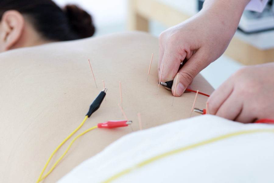
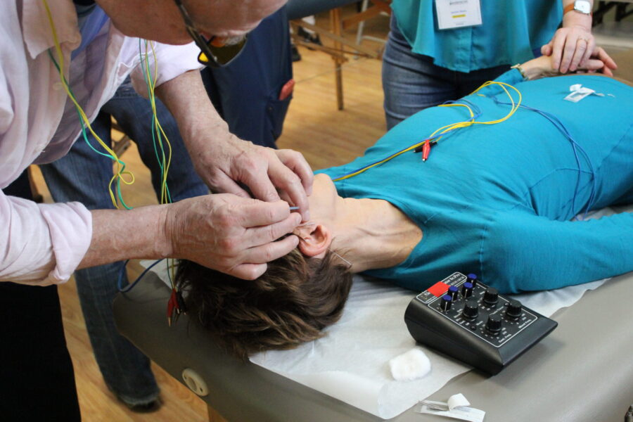
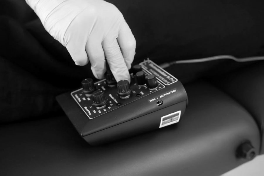
In this study published in the Journal of Electron Spectroscopy and Related Phenomena researchers used state-of-the-art imaging techniques, allowing for improved images of soft tissues, which lead to this important discovery. Here is an excerpt of the article:
Acupuncture points have a higher density of micro-vessels and contain a large amount of involuted microvascular structures. The non-acupuncture points did not exhibit these properties.
The acupuncture points ST36 (Zusanli) and ST37 (Shangjuxu) were shown to have very distinct structural differences than surrounding areas. At the acupuncture points, microvascular densities with bifurcations “can be clearly seen around thick blood vessels” but non-acupuncture point areas showed few thick blood vessels and none showed fine, high density structures. The acupuncture points contained fine structures with more large blood vessels that are several dozen micrometers in size plus beds of high density vascularization of vessels 15-50 micrometers in size. This structure was not found in non-acupuncture point areas.
The researchers note that the size of an acupuncture point “can be estimated by the diameter of microvascular aggregations….” They also commented that other research has found unique structures of acupuncture points and acupuncture meridians using MRI (magnetic resonance imaging), infrared imaging, LCD thermal photography, ultrasound and other CT imaging methods. The researchers commented that many studies using these technological approaches have already shown that acupuncture points exist. They note that “the high brightness, wide spectrum, high collimation, polarization and pulsed structure of synchrotron radiation” facilitated their discovery. They concluded, “Our results demonstrated again the existence of acupoints, and also show that the acupoints are special points in mammals.”